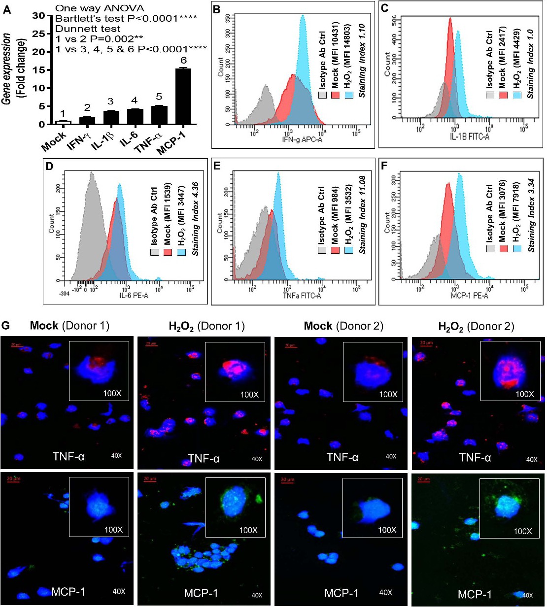
Fig. 5. Oxidative stress promotes the expression of proinflammatory cytokines/chemokine. PBMC were treated with H2O2 for induction of oxidative stress or with vehicle (mock). Total RNA was extracted and IFN-γ, IL-1β, IL-6, TNF-α, and MCP-1 gene expression was determined using real-time RT-PCR as described in materials and methods. At the same time, harvested cells were also stained intracellularly to assess protein expression of these cytokines/chemokine by flow cytometry (SI) and/or confocal microscopy. (A) The data (mean±SEM) obtained from 5 independent determinations with similar results show that H2O2 treatment promoted mRNA expression of IFN-γ (2.0±0.21 fold), IL-1β (3.81±0.02 fold), IL-6 (4.23±0.04 fold), TNF-α (5.10±0.18 fold), and MCP-1 (15.47±0.22 fold) compared to mock. As expected, oxidative stress also enhanced protein expression of (B) IFN-γ (SI=1.10), (C) IL-1β (SI=1.0), (D) IL-6 (SI=4.36), (E) TNF-α (SI=11.08), and (F) MCP-1 (SI=3.34) compared to mock. (G) Confocal microscopy images (40× magnification; inset at 100× magnification) show the elevated TNF-α and MCP-1 expression in PBMC treated with H2O2 as compared to mock, two donors each.